This in vivo µCT system located in the animal facilities at Dept. of Biomedicine, Aarhus University, enables scanning of live animals such as mice and rats with an image resolution of about 10 µm. This means that bone microstructure may be followed over time to enable quantification of for example bone formed and removed during bone (re)modeling and the impact of growth, intervention, or disease in vivo. Moreover, the scanner allows for scanning of soft tissue by use of contrast agents as well as larger human bones in vitro. It thus bridges the information accessible by clinical High Resolution peripheral Quantitative CT (HR-pQCT) and that accessible by high-resolution X-ray microscopy and at synchrotrons, thus ensuring a continuum in the clinical and pre-clinical investigations.
The primary use of the in vivo µCT system will be for studies of the musculoskeletal system in health and disease, but the possibilities provided by VivaCT 80 can be favorably applied much broader and we are happy to assist in projects ranging from biology to materials science, geoscience and beyond. If you have questions or want to know more about the possibilities, you are welcome to contact us.
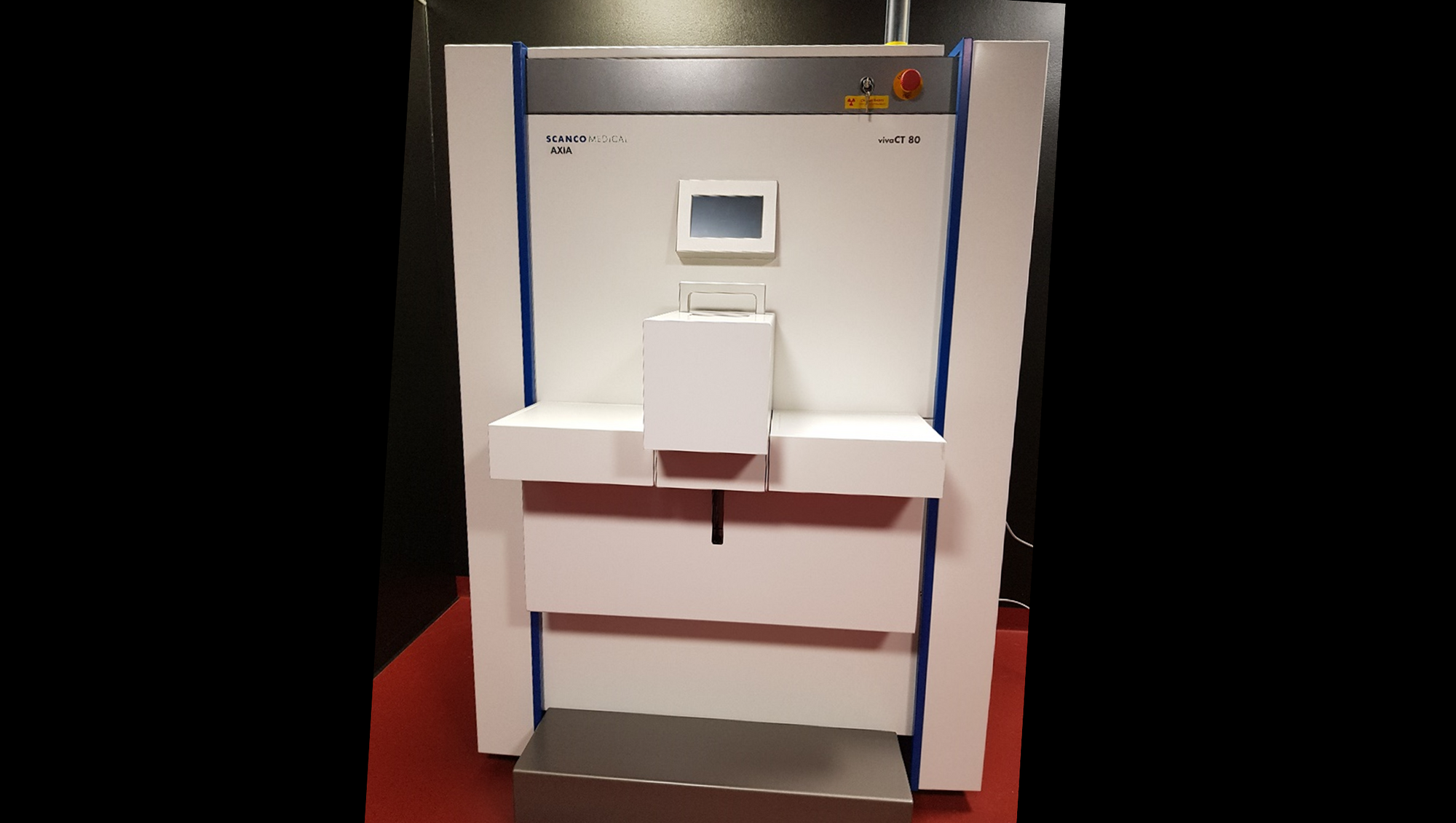
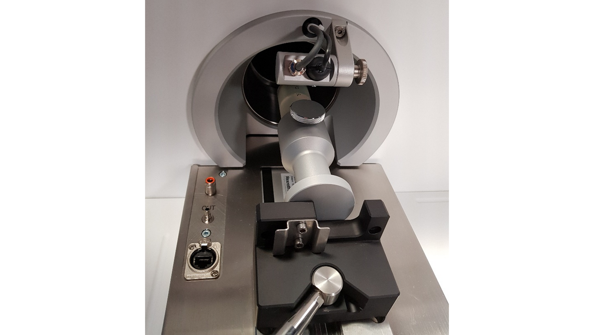

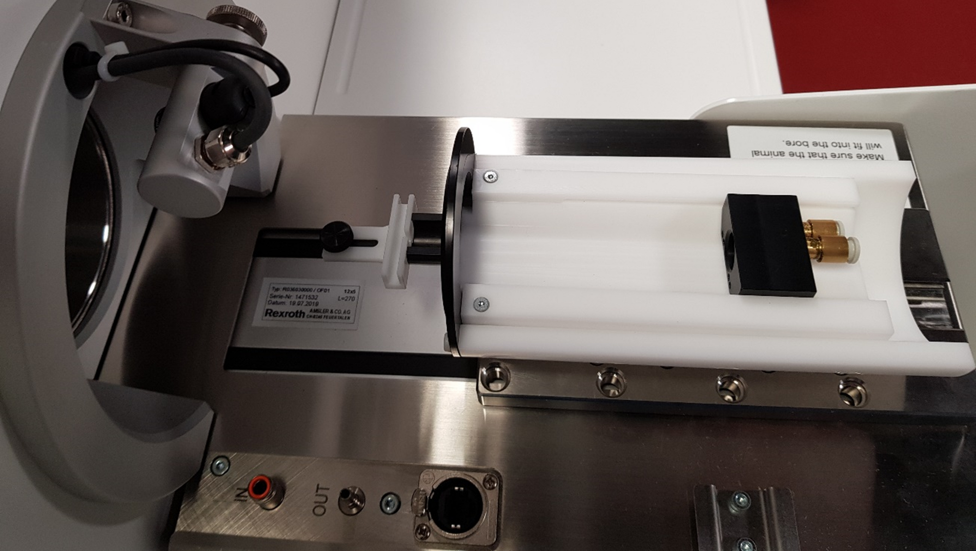
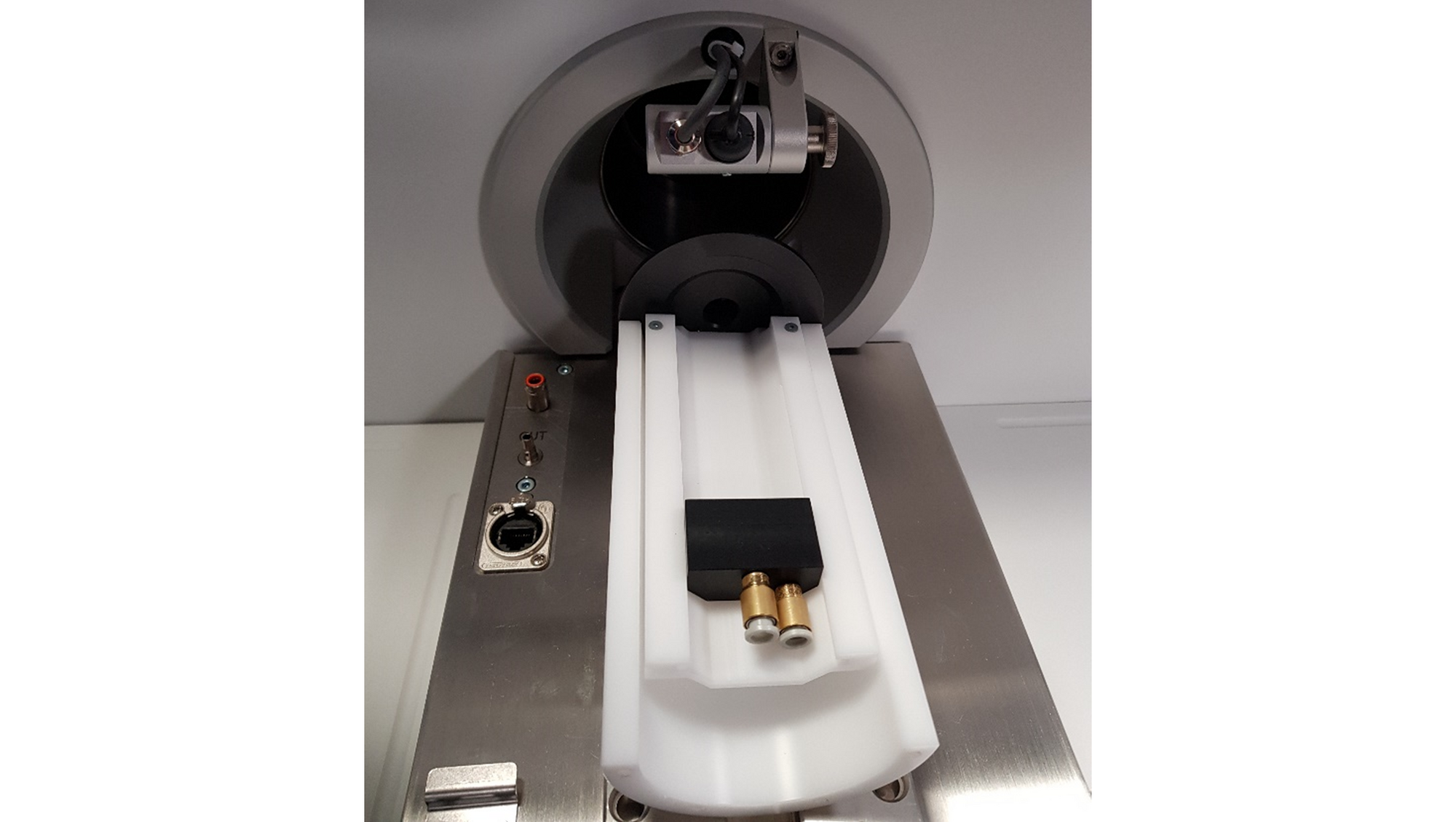
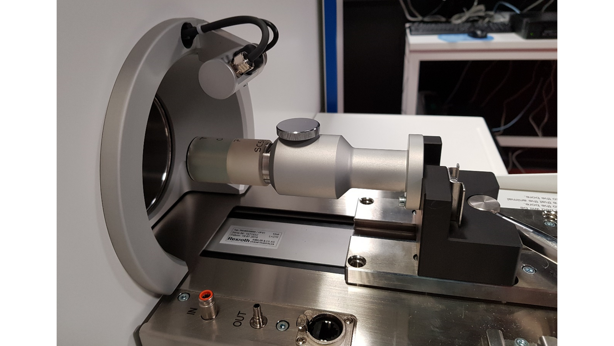
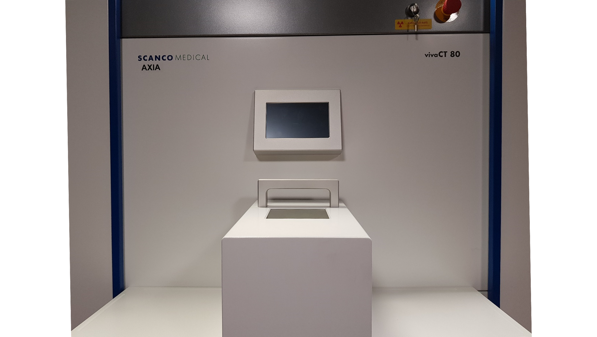
Photos: Jesper Skovhus Thomsen
