Specialized iNANO Lecture: Advances in Quantitative and Three-Dimensional Mapping of Soft Matter by Force Microscopy
Professor Ricardo Garcia, Instituto de Ciencia de Materiales de Madrid, CSIC, Madrid, Spain
Info about event
Time
Location
iNANO Auditorium, Gustav Wieds Vej 14, 8000 Aarhus C

Professor Ricardo Garcia, Instituto de Ciencia de Materiales de Madrid, CSIC, Madrid, SpainAdvances in Quantitative and Three-Dimensional Mapping of Soft Matter by Force MicroscopyProbe microscopy is considered the second most relevant advance in Materials Science since 1960 because its versatility. It is a high spatial resolution microscopy but, among other applications, it could also be transformed into a nanolithography tool. Despite the success of AFM, the technique currently faces limitations in terms of three-dimensional imaging, spatial resolution, quantitative measurements and data acquisition times. Atomic and molecular resolution imaging in air, liquid or ultrahigh vacuum is arguably the most striking feature of the instrument. However, high resolution imaging is a property that depends on both the sensitivity and resolution of the microscope and on the mechanical properties of the material under study. Molecular resolution images of soft matter are hard to achieve. In fact, no comparable high resolution images have been reported for very soft materials such as those with an effective elastic modulus below 10 MPa (isolated proteins, cells, some polymers). Similarly, it is hard to combine the exquisite force sensitivity of force spectroscopy with molecular resolution imaging. Simultaneous high spatial resolution and material properties mapping is still challenging. This presentation reviews some of the above limitations and some recent developments based on the bimodal operation of the AFM to address and overcome them.
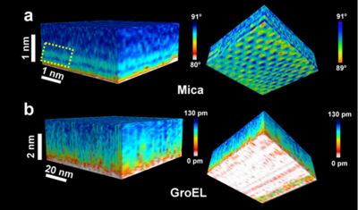
Bimodal AFM 3D images of solid-water volumes. a, 3D map of a mica-water interface. The side view shows variations of the phase shift of mode 2. The stripes are associated to the presence of hydration layers. The view from the mica surface upwards (left panel) shows variations of ϕ2. b, 3D map of a GroEL patch-water interface. The side view shows a slightly rough landscape with variations of the amplitude of about 1 nm. Those variations are interpreted as perturbations in the interface. The view from the GroEL patch upwards shows the relatively rough surface of the GroEL patches. Recent references:
| |
| Host: Associate professor Mingdong Dong, Interdisciplinary Nanoscience Center, Aarhus C, Denmark |

