Facility manager Maja Østergaard and colleagues just published a research paper in Bone highlighting the effect of X-ray microcomputed tomography scan settings on osteocyte lacunar form using AXIA infrastructure.
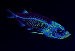
AXIA-tomogram "Like a fish out of water" won first price in this year's Science as Art competition at iNANO.
The tomogram was acquired with AXIA's 3D X-ray microscope and visualized in Dragonfly 2022.1 (ORS) using a discrete version of the Rainbow lookup table from a transparent purple (flesh and fish scales) through blue and green (skeleton) to an opaque red for increasingly dense material.
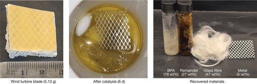
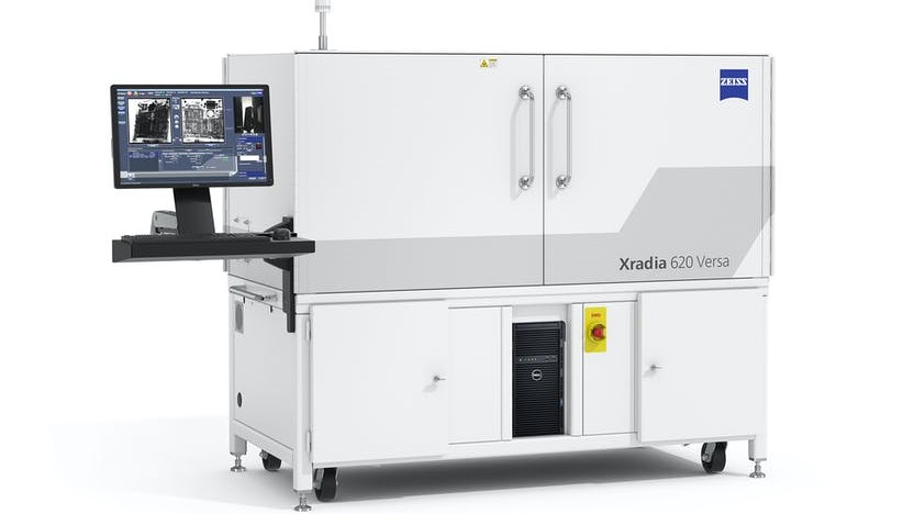
We have been so fortunate to work with researchers from the Skrydstrup group and Danish Technological Institute who have developed a chemical process to extract virgin-grade materials from wind turbine blades in one process. The quality of the extracted glass fibres was assessed using our Xradia 620 Versa microscope among other exciting techniques. The work has been published in nature.
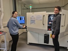
This fall we held a course on X-ray computed tomography in practice, and from that we now have the first independent users of our 3D X-ray microscope.
In the course, the students were taught the basic principles of computed tomography followed by four days of experimental and computer exercises to learn the instrumentation as well as simple tools for data analysis.
We look forward to repeating the success next year!
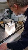

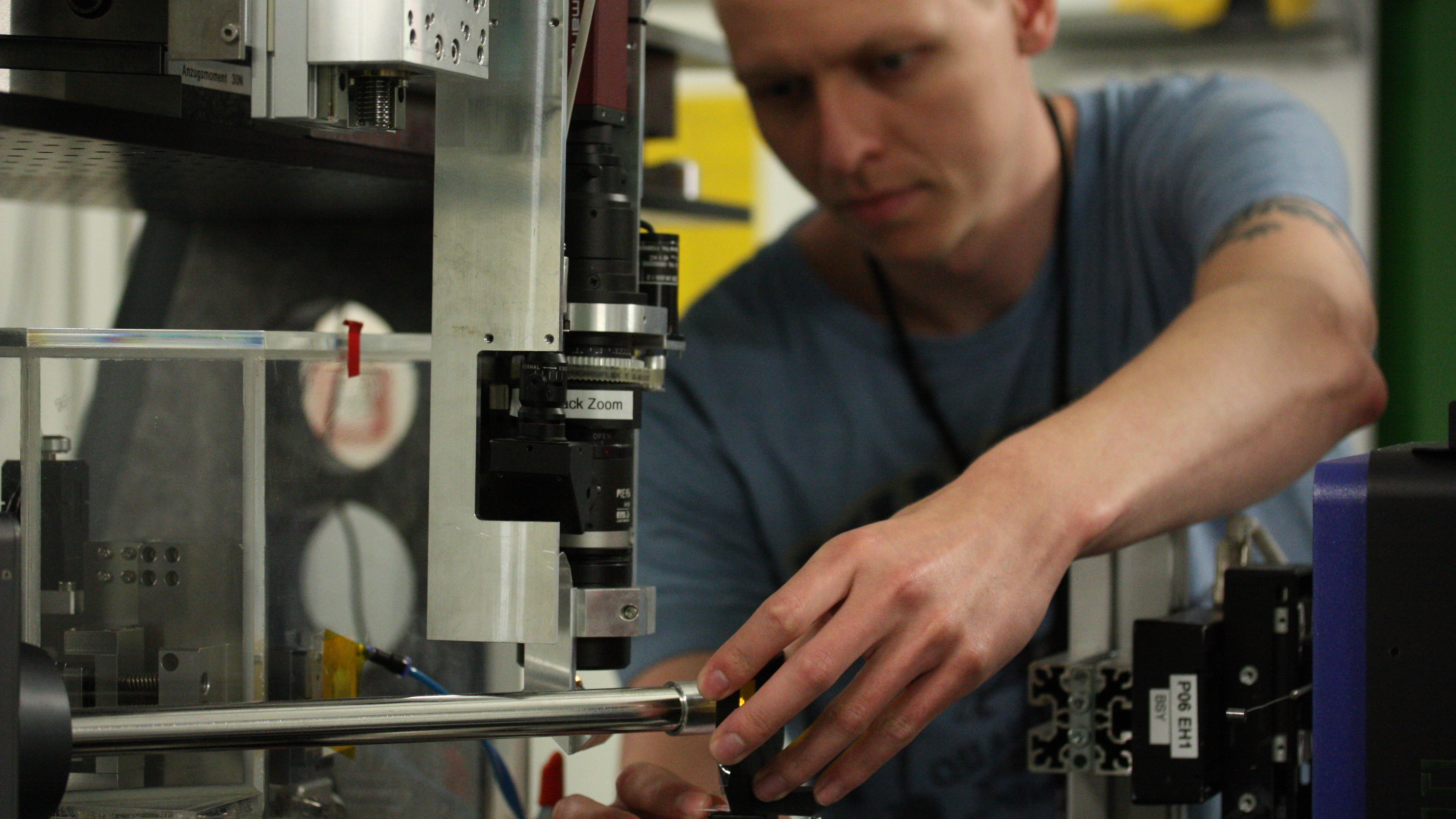
Inspired by the system published by Holler et al, AC-TAP Carsten Pedersen has build a lathe for milling of small cylindric samples (µm-mm) optimized for tomography measurements.
The system is already being used efficiently, most recently in combination with Ga ion fibbing guided by µCT images to produce ultra-small samples for a synchrotron experiment currently being conducted at the NanoMAX beamline at MAX-IV in Lund.