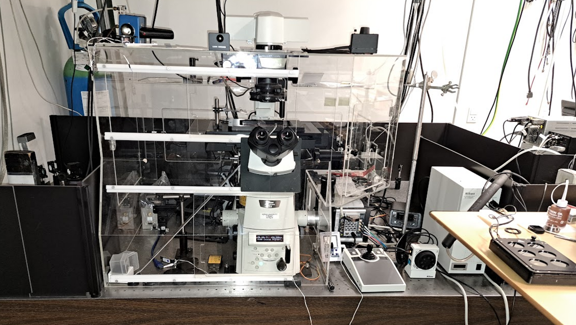
The Nikon Structured Illumination Microscope (N-SIM) allows for fluorescence imaging with a two-fold better spatial resolution than diffraction limited traditional fluorescence microscopy. SIM is well suited for biological imaging of small cellular structures and our setup is equipped with imaging software featuring user-friendly functionalities for easy design of image recordings and reconstruction of the obtained image sequences. Our single- and multi-well plate stagetop incubators offer researchers the opportunity to study cells (and other specimens) under controlled atmospheric conditions and various temperatures.
Our setup is also equipped with a fiber spectrometer, which can record fluorescence spectra on the sample stage. This can be useful for observing spectral changes of a dye due to environmental shifts in cells or solution.
If you are interested or have additional questions, please contact us.
Cellular imaging with sub-diffraction resolution. See resolution below.
| 3D SIM | 2D SIM | TIRF SIM | |
|---|---|---|---|
| x/y | 115 nm | 90 nm | 90 nm |
| z | 269 nm | 220 nm | 220 nm |

Up to 50 µm deep
Up to 3 µm deep
For objects very close to the coverslip surface
Other possibilities are also available upon request. Please contact us to hear more.
