Atomic force microscopy (AFM) is a scanning probe technique, in which a sharp tip, which is positioned on a cantilever, is scanned in a raster-pattern along a surface. Due to forces acting between the surface and tip the cantilever is deflected away from its equilibrium position. It is possible to record the deflection as function of time and, thereby, form an image of the surface topography with close to atomic resolution.
The relevant forces between the tip and the surface are:
The Van der Waals force between two atoms shows up due to fluctuations in the charge-density of the atomic cores and the surrounding electrons. Although the force is typically perceived as a force between single atoms, the force also plays an important role in the interaction of macroscopic objects. In this case the force depends on the specific geometry of the objects.
The Coulumb force is the force between charged particles or objects.
The meniscus force is the force that arise due to surface tension in a water-meniscus, which spans and connects tip and surface. This meniscus is present when scanning in gas-phase as well as in vacuum with sufficiently small tip-surface distance. The amplitude of the force depends on the two curvatures of the meniscus, which again depends on tip radius.
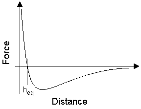
The force vs. distance curve is shown in Figure1 below. At distances larger than heq the effective force is attractive, while in the region (0,heq) the force is dominated by the strongly repulsive Van der Waals force (the VdW-force follows an r-12-law in this range). This repulsive range is also called the contact range.

Figure1.
The distance between the surface and the tip as well as the horizontal movement is controlled by an arrangement of piezoelectric elements controlling the x,y (horizontal), and z (vertical) coordinates of the sample surface. The angle of deflection of the cantilever is measured as function of time using a laserbeam reflecting off the cantilever and into a two-segment photodiode.
An illustration of how the reflecting laser beam method measures deflection.
The topographical data are equal to the sum of the deflection and the z-position of the surface. By using a feedback system the piezoelectric elements can be controlled in the z-direction, so that the deflection of the cantilever is at a minimum. In this case, the topographical data is approximately equal to the z-position of the surface only. The advantage of using feedback is the limited amount of force being excerted on the surface, which makes the AFM-technique more gentle to soft biological molecules and particles.
Even feedback may not be enough when imaging really soft, fragile or very lightly bound material, which is easily pushed away by the cantilever. Other more gentle modes of AFM have, however, been invented, e.g., tapping mode where the tip is not 'scraped' along the surface, as is the case for the described contact mode, but 'tapped' along the surface.
Besides acquisition of topographical data, AFM is used to measure the friction between the tip and surface. When scanning in contact-mode any hindrance in the path of the tip will induce torsion in the cantilever. This torsion can be measured by the reflecting laser beam method and can subsequently be converted into an image. The friction measured by this method clearly does not correspond to the classical macroscopic concept of friction. Macroscopic friction effectively corresponds to a very large number of tips, which simultaneously interact with a surface consisting of a very large number of bumps. This 'microscopic' friction-measurement is the most local measurement of friction attainable.
Aarhus University is currently operating several AFM systems, including the Bruker Agilent.
iNANO's current research using AFM is wide-ranging and includes: surface characterisation, measurements of binding forces of biomolecules to surfaces, and imaging of several different species of biomolecules on different types of surfaces.
Typically, iNANO would like to know the behaviour and properties of specific molecules on specific surfaces, e.g., the conformation of molecules (proteins, DNA, RNA etc.), clustering (as in the case of e.g. fibrinogen), and self-organization.
Examples of research and images will be published on this site very soon...